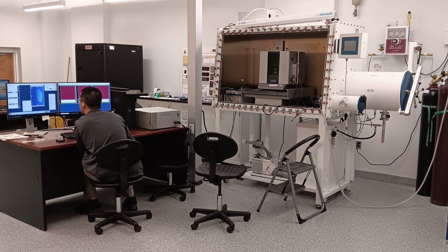
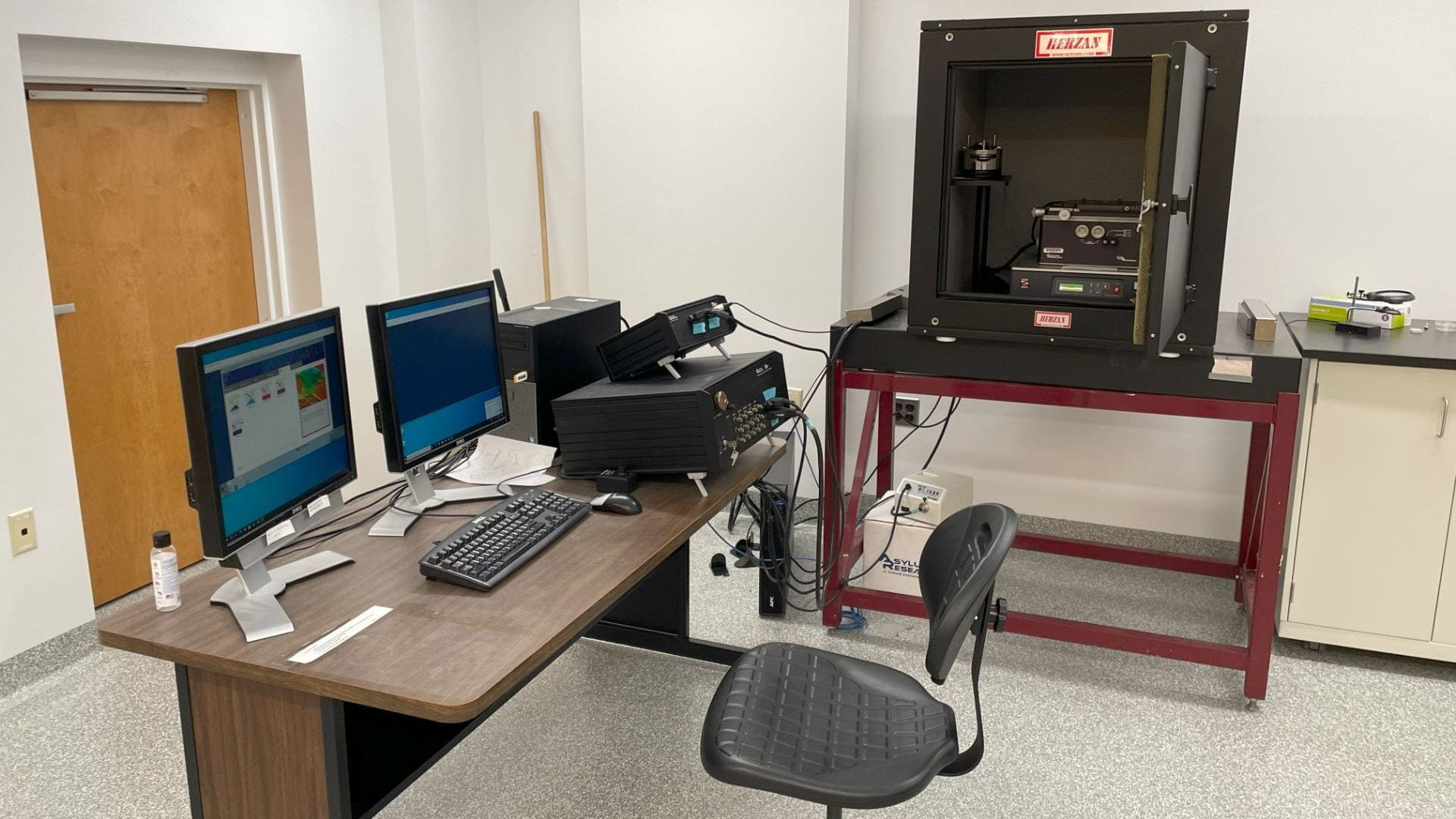
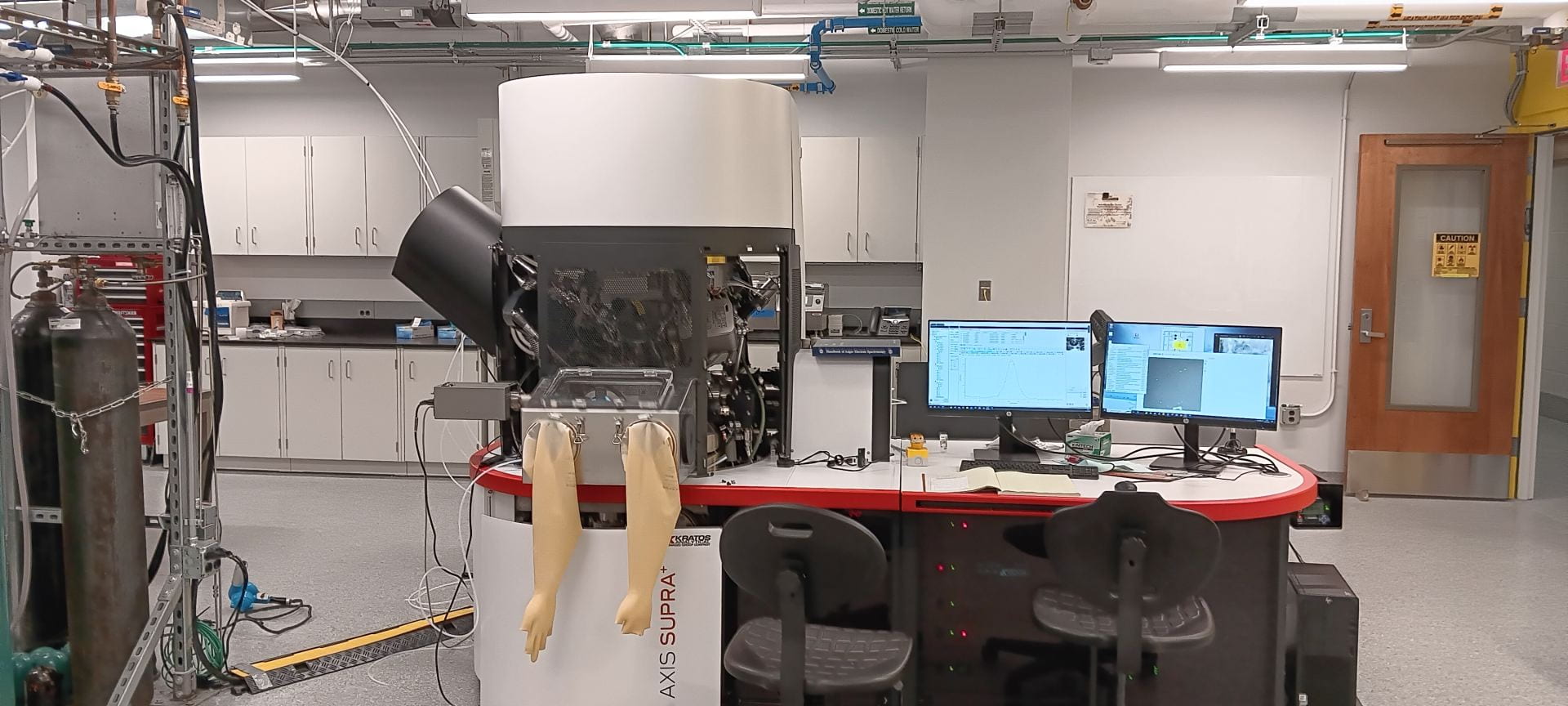
The Surface Analysis center houses two Atomic Force Microscopes:
Both internal and external users can either submit samples for analysis (full service) or be trained to use the AFM as an independent user.
AFM obtains high-resolution images and various material properties at the nanoscale for polymers, thin films, devices, advanced materials and biological samples amongst others, Z resolution is on the tens of picometer scale.
More details about each of our instruments and their capabilities can be found below.
Cypher ES™ Environmental AFM

This is the most state of art AFM in SAC. The instrument can provide controlled gas/liquid/humidity environment. It’s also capable of heating (250 ℃), cooling-heating (0~120℃) and high voltage (+/- 220V).
Sample size requirement
Sample size:
X,Y: Must fit within a 15mm diameter disk
Z: 8mm thickness maximum
Sample surface roughness:
3 μm maximum (bottom of valley to top of mountain)
Instrumental details:
Max scan size: X,Y 30 μm
Environmental options:
- Liquid- regular
- Liquid- perfusion
- Humidity controlled
- Heating (Room temperature to 250℃)
- Cooling-heating (0~120℃)
Imaging mode:
- Contact topography (Lateral Force Mode)
- Tapping Mode with Q-control
- BlueDrive™ – High speed imaging 10x-20x faster
- Force Modulation
- Frequency Modulation
- Phase imaging
- Force Curve Mode
- Fast Force Mapping (Force volume)
- Bimodal Dual AC™
- Magnetic Force Microscopy (MFM)
- Electrostatic Force Microscopy (EFM)
- Scanning Kelvin Probe Microscopy (SKPM)
- MicroAngelo™ (Nanolithography and nanomanipulation)
- Electrochemical AFM (EC-AFM)
- AM-FM Viscoelastic Mapping Mode
- AM-FM/Loss Tangent
- Conductive AFM
- Dual gain ORCA™
- Piezoresponse force microscopy (PFM)
MFP-3D Origin™ AFM

This instrument is suitable for much larger samples. It also has a variety of scanning modes.
Sample submission size requirement
Sample size:
X,Y: 80mm x80mm maximum
Z: 8mm thickness maximum
Sample surface roughness:
6 μm maximum (bottom of valley to top of mountain)
Instrumental details:
Max scan size: X,Y 90μm
Imaging mode:
- Contact topography (Lateral Force Mode)
- Tapping Mode with Q-control
- Force Modulation
- Frequency Modulation
- Phase Imaging
- Force Curve Mode
- Force Mapping (Force Volume)
- Bimodal Dual AC™
- Magnetic Force Microscopy (MFM)
- Electrostatic Force Microscopy (EFM)
- MicroAngelo™ (Nanolithography and nanomanipulation)
