Once you have appropriately exported your data to CASA XPS;
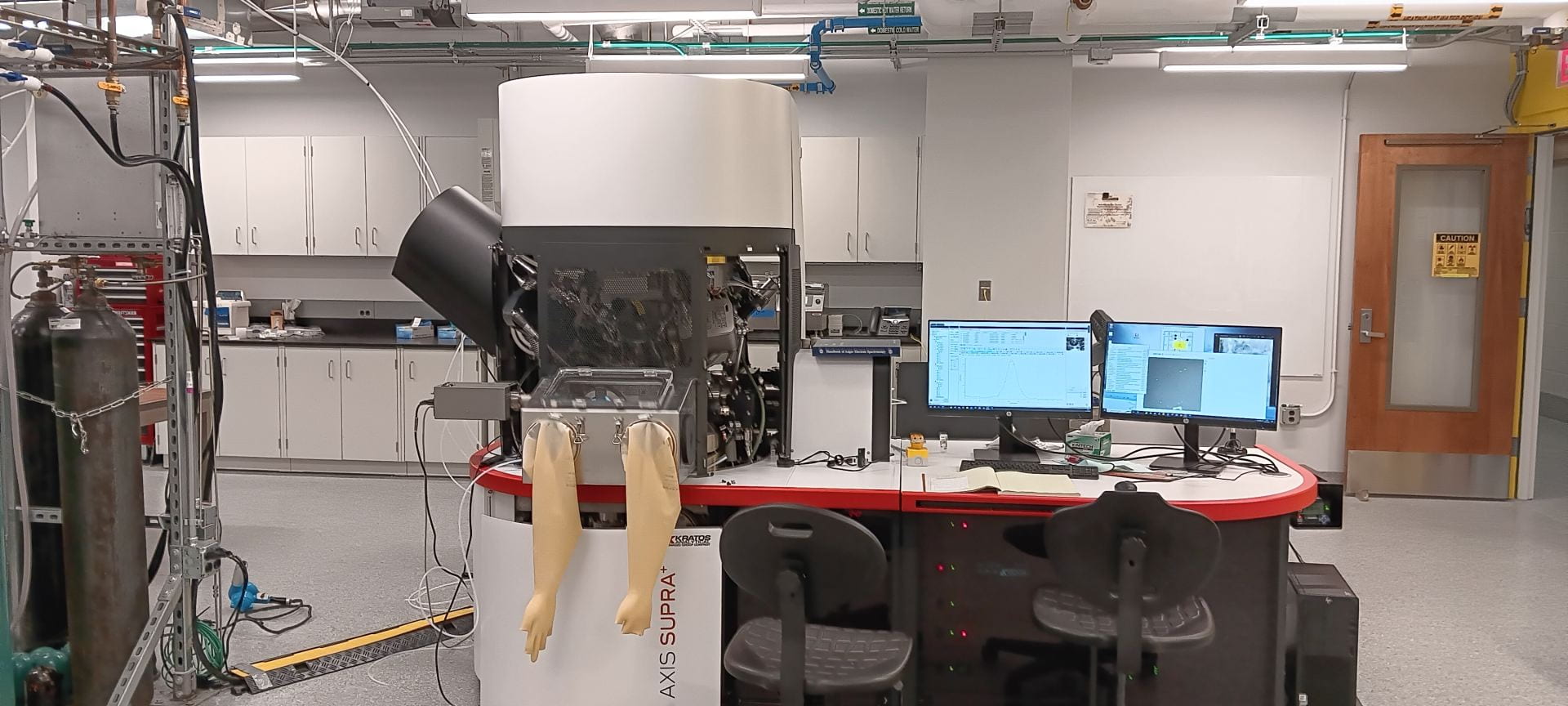
- For accurate quantification you must first ensure you are using the correct relative sensitivity factors and appropriate intensity calibration for your specific XPS instrument and x-ray source energy. Using the default RSF’s and Intensity calibration settings in CASA will lead to incorrect quantification. Before starting data analysis ensure you have loaded the correct library of relative sensitivity factors. In our case, for the Kratos Axis instruments follow the procedure below.
- Open the elements window which can be found under the options tab
- Select the input file tab in the element library window
- Press “Browse Lib Dir…” button
- select “casaXPS_KratosAxis-F1s…” from the list, then press select
- Back in the element Library window press load at the bottom of the window
- If you have followed the above procedure correctly you should find that the RSF assigned to C 1s is 0.278.
- For the Intensity calibration ensure that in the wide and narrow quantify windows (under options) have the check box checked for automatic calibration , the adjacent box says zero and underneath is written “Escape D.C.: KE Exp. / ADC Magic Angle”. These settings reflect the fact that the transmission file is automatically included with the raw data when it is exported from the escape software and imported into CASA.
- Next use CASAXPS to identify and label all your expected and unexpected peaks in the survey spectrum. By doing this first you will identify any peak overlaps you need to take into account when analyzing the high resolution spectra. Do not assume that all peak structure in a high resolution spectrum are due to the element of interest, there are often peaks that overlap from unexpected or expected elements or background structure.
- Next use the Regions tab in the quantification parameters window to create a region for each of your high resolution spectra assuming there are no overlaps. There are multiple backgrounds to choose from, the Shirley background, which works well in the majority of cases. Once you have chosen a background use the same one for all regions in the dataset. Make sure to extend the back ground at least 5 – 10 data points before and after the peak, and that your background is centered in any noise.
- Before going any further you will need to calibrate your energy scale, ie reference your data to a peak with a known binding energy. Referencing to the adventitious hydrocarbon peak at 284.8 eV is commonly used, however it is not always the best choice, other choices may be an intense narrow O2- peak of an oxide or a highly electronegative peak like F 1s in a fluoride for example.
- You can now display the relative percentage atomic concentrations of highlighted regions using the quantification tab in the annotation window.
- If you need to go further and attempt some peak fitting, first determine whether you may have differential sample charging, if you do, do not attempt peak fitting as it will lead to incorrect analysis and conclusions, you should rerun the samples and take measures to avoid differential sample charging.
- Peak fitting must be done very carefully, peak fitting requires understanding both the physical principles of the experiment as well as chemistry knowledge about your material under study, it is not black and white and most often grey! Peak fitting should not violate any physical facts (discussed below) and should make chemical sense, a peak fit that fits data reasonably well, while making physical and chemical sense, is much better than a peak fit that fits the data envelope perfectly but neglects physical and chemical sense. There are a staggering number of analysis errors in literature due to incorrect peak fitting. Please adhere to the following facts when peak fitting;
- FWHM (full with at half maximum)
- Peaks cannot be narrower than the x-ray line width (~0.25 eV for the monochromatic Al x-rays). Peaks are additionally broadened by instrumental broadening (Gaussian) as well as the natural linewidth of your energy level (Lorentzian). The smallest linewidth measured with an instrument using Al monochromatic x-rays is 0.32 eV for Re 4f7/2. It is therefore not physically possible for any peak to be narrower than 0.32 eV.
- Most peaks will fall between ~0.5 – 2.0 eV. Pure metal peaks tend to have the narrowest FWHM’s (~0.5 – 1.0 eV), followed by inorganic compounds (~0.5-1.5 eV) with organic compounds tending to have the largest FWHM’s (~1.0-2.0 eV or larger)
- Spin orbit split components tend to have the same FWHM’s as one another, but their are exceptions for example Ti 2p.
- FWHM constraints should be used routinely for peak fitting, FWHM’s often only differ by 10% from one another, it is often a good approximation to fix FWHM’s to be the same as one another that would be expected to be similar. Covalent compounds tend to give broader XPS peaks than ionic compounds. Metals usually have the narrowest peaks. For example when fitting the C 1s/ O1s/ N 1s etc of organic compounds a more meaningful peak fit can be obtained by fixing all the FWHMs to be the same in a given region rather than letting them vary, this will also allow more accurate comparison while comparing very similar materials.
- Sample charging during data collection can cause peak broadening sometimes up to 3 -4 eV or higher. To minimize or ideally to prevent this the user should pay particular attention when running the sample to optimize the charge neutralizer to give the narrowest FWHM. Simple sample charging can usually be spotted when all the peaks are boarder than expected.
- Peak Shape
- Peak shape is a mix of Gaussian and Lorentzian peak shapes. In CASA XPS using “GL(30)” (Gaussian/Lorentzian product formula) for most peaks works well for data collected on our instruments using the monochromatic Al x-ray source.
- Not all peaks are symmetric, for example conductive samples such as metals, graphite etc. have varying degrees of asymmetry to the higher binding energy side of the peak due to interaction of the outgoing photoelectrons with the electrons in the conduction band, the extent of asymmetry is proportional to the the density of states at the fermi energy. I suggest using the ad hoc exponential tail using “A(a,b,n)GL(p)“ for the asymmetric line shape in CASA as a good choice. It is common for researchers to incorrectly fit these asymmetric tails using multiple symmetrical peaks and incorrectly assigning functionality to these extra peaks. Please be careful!
- Spin-orbit split components
- Typically spin orbit split components have the same FWHM as each other, however there are exceptions such as Ti 2p where the 1/2 component is significantly broader than the 3/2 component, this is a result of Coster-Kronig broadening. It is a good first approximation to constrain you spin-orbit split components to have equal FWHM’s, be on the lookout for exceptions though.
- Area ratios of spin orbit split components are determined by quantum mechanic as follows;
- FWHM (full with at half maximum)
| Subshell | Spin (s) | orbital angular momentum (l) | j values (l+s) | Peak Area Ratio (2j+1) |
| s | +/-1/2 | 0 | 1/2 | N/A |
| p | +/-1/2 | 1 | 3/2, 1/2 | 2:1 |
| d | +/-1/2 | 2 | 5/2, 3/2 | 3:2 |
| f | +/-1/2 | 3 | 7/2, 5/2 | 4:3 |
To be continued work in progress!
